


Figure 2.
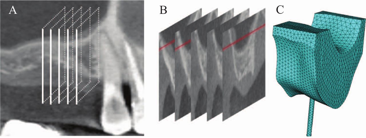
The construction process of a subject-specific model.
A: the computed tomography of the maxillary right molar region; B: five buccolingual section images to create a solid volume; C: meshed three-dimensional FE model with the loading rod.
FE: finite element